Sistem kardiovaskular# Cardiovascular System
Sistem kardiovaskular
Sistem kardiovaskular terdiri dari jantung, pembuluh darah, dan sekitar 5 liter darah yang dibawa oleh pembuluh darah. Bertanggung jawab untuk mengangkut oksigen, nutrisi, hormon, dan produk limbah seluler ke seluruh tubuh, sistem kardiovaskular didukung oleh organ tubuh yang paling sulit - jantung, yang hanya seukuran kepalan tangan tertutup. Bahkan pada saat istirahat, rata-rata jantung dengan mudah memompa lebih dari 5 liter darah ke seluruh tubuh setiap menitnya.
****Anatomi Sistem Kardiovaskular****
****Jantung****
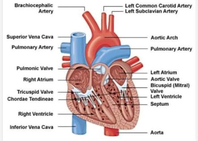
Jantung adalah organ pemompa otot yang terletak di medial ke paru-paru di sepanjang garis tengah tubuh di daerah toraks. Ujung bawah jantung, yang dikenal sebagai puncaknya, diputar ke kiri, sehingga sekitar 2/3 jantung terletak di sisi kiri tubuh dengan 1/3 yang lain di sebelah kanan. Bagian atas jantung, yang dikenal sebagai dasar jantung, terhubung ke pembuluh darah besar tubuh: aorta, vena cava, pulmonary trunk, dan pulmonary veins.
Peredaran Peredaran Darah
Ada 2 loop peredaran primer dalam tubuh manusia: sirkulasi pulmoner dan loop sirkulasi sistemik.
Sirkulasi paru mengangkut darah terdeoksigenasi dari sisi kanan jantung ke paru-paru, di mana darah mengambil oksigen dan kembali ke sisi kiri jantung. Ruang pemompaan jantung yang mendukung sirkulasi pulmoner adalah atrium kanan dan ventrikel kanan.
Sirkulasi sistemik membawa darah yang sangat beroksigen dari sisi kiri jantung ke semua jaringan tubuh (kecuali jantung dan paru-paru). Sirkulasi sistemik menghilangkan limbah dari jaringan tubuh dan mengembalikan darah terdeoksigenasi ke sisi kanan jantung. Atrium kiri dan ventrikel kiri jantung adalah ruang pemompaan untuk sirkulasi sirkulasi sistemik.
****Vena Pembuluh****
Vena Pembuluh darah adalah jalan raya tubuh yang memungkinkan darah mengalir dengan cepat dan efisien dari jantung ke setiap area tubuh dan kembali lagi. Ukuran pembuluh darah sesuai dengan jumlah darah yang melewati pembuluh darah. Semua pembuluh darah mengandung daerah berongga yang disebut lumen dimana darah mengalir. Sekitar lumen adalah dinding pembuluh darah, yang mungkin tipis dalam kasus kapiler atau sangat tebal dalam kasus arteri. Semua pembuluh darah dilapisi dengan lapisan tipis epitel skuamosa sederhana yang dikenal sebagai endotelium yang menjaga sel darah di dalam pembuluh darah dan mencegah gumpalan terbentuk. Lapisan endothelium melapisi seluruh sistem peredaran darah, sampai ke bagian dalam jantung, di mana ia disebut endokardium. Ada tiga tipe utama pembuluh darah: arteri, kapiler dan vena.
Pembuluh darah sering disebut baik di daerah tubuh dimana mereka membawa darah atau ke struktur terdekat. Misalnya, arteri brachiocephalic membawa darah ke daerah brachial (lengan) dan cephalic (kepala). Salah satu cabangnya, arteri subklavia, berjalan di bawah klavikula; maka nama subclavian. Arteri subklavia memasuki daerah aksila dimana ia dikenal sebagai arteri aksilaris.
***Arteri dan Arterioles:**** Arteri adalah pembuluh darah yang membawa darah menjauh dari jantung. Darah yang dibawa oleh arteri biasanya sangat beroksigen, baru saja meninggalkan paru-paru dalam perjalanan menuju jaringan tubuh. Batang pulmonal dan arteri sirkit sirkulasi paru memberikan pengecualian terhadap peraturan ini - arteri ini membawa darah terdeoksigenasi dari jantung ke paru-paru agar beroksigen.
Arteri menghadapi tekanan darah tingkat tinggi saat membawa darah didorong dari jantung dengan kekuatan besar. Untuk menahan tekanan ini, dinding arteri lebih tebal, lebih elastis, dan lebih berotot dibandingkan dengan pembuluh darah lainnya. Arteri terbesar dari tubuh mengandung persentase jaringan elastis yang tinggi yang memungkinkan mereka meregangkan dan mengakomodasi tekanan jantung.
Arteri yang lebih kecil lebih berotot dalam struktur dindingnya. Otot halus dinding arteri dari arteri yang lebih kecil ini berkontraksi atau meluas untuk mengatur aliran darah melalui lumennya. Dengan cara ini, tubuh mengontrol berapa banyak darah mengalir ke berbagai bagian tubuh dalam situasi yang bervariasi. Pengaturan aliran darah juga mempengaruhi tekanan darah, karena arteri yang lebih kecil memberi area kurang darah untuk mengalir dan oleh karena itu meningkatkan tekanan darah di dinding arteri.
Arterioles adalah arteri yang sempit yang bercabang dari ujung arteri dan membawa darah ke kapiler. Mereka menghadapi tekanan darah jauh lebih rendah daripada arteri karena jumlah mereka yang lebih besar, penurunan volume darah, dan jarak dari tekanan jantung langsung. Dengan demikian dinding arteriol jauh lebih tipis dibanding arteri. Arterioles, seperti arteri, mampu menggunakan otot polos untuk mengendalikan aperture dan mengatur aliran darah dan tekanan darah.
***Kapiler: ***Kapiler adalah pembuluh darah terkecil dan tertipis di dalam tubuh dan juga yang paling umum. Mereka dapat ditemukan berjalan di hampir setiap jaringan tubuh dan membatasi tepi jaringan avaskular tubuh. Kapiler terhubung ke arteriola pada satu ujung dan venula di sisi lain.
Kapiler membawa darah sangat dekat dengan sel-sel jaringan tubuh untuk menukar gas, nutrisi, dan produk limbah. Dinding kapiler hanya terdiri dari lapisan tipis endotelium sehingga ada jumlah minimum struktur yang mungkin antara darah dan jaringan. Endothelium bertindak sebagai filter untuk menjaga sel darah di dalam pembuluh darah sementara membiarkan cairan, gas terlarut, dan bahan kimia lainnya menyebar sepanjang konsentrasi gradien ke atau keluar dari jaringan.
Sphincters praprofi adalah pita otot polos yang ditemukan di ujung arteriol kapiler. Saraf ini mengatur aliran darah ke kapiler. Karena ada persediaan darah yang terbatas, dan tidak semua jaringan memiliki kebutuhan energi dan oksigen yang sama, sphincters precapillary mengurangi aliran darah ke jaringan yang tidak aktif dan membiarkan aliran bebas ke jaringan aktif.
Vena dan Venules:Vena adalah pembuluh balik tubuh yang besar dan berfungsi sebagai pembawa kembali arteri darah. Karena arteri, arteriol, dan kapiler menyerap sebagian besar kekuatan kontraksi jantung, vena dan venula mengalami tekanan darah yang sangat rendah. Kurangnya tekanan ini memungkinkan dinding pembuluh darah menjadi lebih tipis, kurang elastis, dan kurang berotot dibanding dinding arteri.
Vena bergantung pada gravitasi, inersia, dan kekuatan kontraksi otot rangka untuk membantu mendorong darah kembali ke jantung. Untuk memudahkan pergerakan darah, beberapa pembuluh darah mengandung banyak katup satu arah yang mencegah darah mengalir menjauh dari jantung. Sebagai otot rangka dalam tubuh berkontraksi, mereka memeras vena terdekat dan mendorong darah melewati katup yang lebih dekat ke jantung.
Saat otot mengendur, katup menjebak darah sampai kontraksi lain mendorong darah mendekati jantung. Venules mirip dengan arterioles karena merupakan pembuluh darah kecil yang menghubungkan kapiler, tapi tidak seperti arteriol, venula terhubung ke pembuluh darah dan bukan arteri. Venips mengambil darah dari banyak kapiler dan menyimpannya ke pembuluh darah yang lebih besar untuk dibawa kembali ke jantung.
****Sirkulasi Koroner****
Hati memiliki kumpulan pembuluh darah sendiri yang memberi miokardium oksigen dan nutrisi yang diperlukan untuk memompa darah ke seluruh tubuh. Arteri koroner kiri dan kanan bercabang dari aorta dan memberikan darah ke sisi kiri dan kanan jantung. Sinus koroner adalah vena pada sisi posterior jantung yang mengembalikan darah terdeoksigenasi dari miokardium ke vena cava.
****Sirkulasi Portal Hepatik****
Vena lambung dan usus melakukan fungsi unik: alih-alih membawa darah langsung kembali ke jantung, mereka membawa darah ke hati melalui vena portal hepatik. Darah yang meninggalkan organ pencernaan ini kaya nutrisi dan bahan kimia lain yang diserap dari makanan. Hati menghilangkan racun, menyimpan gula, dan memproses produk pencernaan sebelum mencapai jaringan tubuh lainnya. Darah dari hati kemudian kembali ke jantung melalui vena cava inferior.
****Darah****
Rata-rata tubuh manusia mengandung sekitar 4 sampai 5 liter darah. Sebagai jaringan ikat cairan, ia mengangkut banyak zat melalui tubuh dan membantu menjaga homeostasis nutrisi, limbah, dan gas. Darah terdiri dari sel darah merah, sel darah putih, trombosit, dan cairan plasma.
Engglish
Cardiovascular System
The cardiovascular system consists of the heart, blood vessels, and the approximately 5 liters of blood that the blood vessels transport. Responsible for transporting oxygen, nutrients, hormones, and cellular waste products throughout the body, the cardiovascular system is powered by the body's hardest-working organ - the heart, which is only about the size of a closed fist. Even at rest, the average heart easily pumps over 5 liters of blood throughout the body every minute.
Anatomy of the Cardiovascular System The heart of
the heart is a muscle-pumping organ located medial to the lungs along the midline of the body in the thoracic region. The lower end of the heart, known as the apex, is turned left, so that about 2/3 of the heart is located on the left side of the body with the other 1/3 on the right. The top of the heart, known as the base of the heart, connects to the large blood vessels of the body: the aorta, the vena cava, the pulmonary trunk, and the pulmonary veins. Circulatory Circulation
There are 2 primary circulating loops in the human body: the pulmonary circulation and the loop of the systemic circulation. deoxygenated blood from the right side of the heart to the lungs, where the blood takes oxygen and returns to the left side of the heart.
The heart pumping chamber that supports pulmonary circulation is the right atrium and the right ventricle. Systemic circulation carries very oxygenated blood from the left side of the heart to all body tissues (except the heart and lungs).
Systemic circulation removes waste from body tissues and restores deoxygenated blood to the right side of the heart. The left atrium and the left ventricle of the heart are the pumping chambers for the circulation of the systemic circulation.
****Blood Vessels****
Blood vessels are the body’s highways that allow blood to flow quickly and efficiently from the heart to every region of the body and back again. The size of blood vessels corresponds with the amount of blood that passes through the vessel. All blood vessels contain a hollow area called the lumen through which blood is able to flow. Around the lumen is the wall of the vessel, which may be thin in the case of capillaries or very thick in the case of arteries.
All blood vessels are lined with a thin layer of simple squamous epithelium known as the endothelium that keeps blood cells inside of the blood vessels and prevents clots from forming. The endothelium lines the entire circulatory system, all the way to the interior of the heart, where it is called the endocardium.
There are three major types of blood vessels: arteries, capillaries and veins. Blood vessels are often named after either the region of the body through which they carry blood or for nearby structures. For example, the brachiocephalic artery carries blood into the brachial (arm) and cephalic (head) regions. One of its branches, the subclavian artery, runs under the clavicle; hence the name subclavian. The subclavian artery runs into the axillary region where it becomes known as the axillary artery.
Arteries and Arterioles: Arteries are blood vessels that carry blood away from the heart. Blood carried by arteries is usually highly oxygenated, having just left the lungs on its way to the body’s tissues. The pulmonary trunk and arteries of the pulmonary circulation loop provide an exception to this rule – these arteries carry deoxygenated blood from the heart to the lungs to be oxygenated.
Arteries face high levels of blood pressure as they carry blood being pushed from the heart under great force. To withstand this pressure, the walls of the arteries are thicker, more elastic, and more muscular than those of other vessels. The largest arteries of the body contain a high percentage of elastic tissue that allows them to stretch and accommodate the pressure of the heart.
Smaller arteries are more muscular in the structure of their walls. The smooth muscles of the arterial walls of these smaller arteries contract or expand to regulate the flow of blood through their lumen. In this way, the body controls how much blood flows to different parts of the body under varying circumstances. The regulation of blood flow also affects blood pressure, as smaller arteries give blood less area to flow through and therefore increases the pressure of the blood on arterial walls.
Arterioles are narrower arteries that branch off from the ends of arteries and carry blood to capillaries. They face much lower blood pressures than arteries due to their greater number, decreased blood volume, and distance from the direct pressure of the heart. Thus arteriole walls are much thinner than those of arteries. Arterioles, like arteries, are able to use smooth muscle to control their aperture and regulate blood flow and blood pressure.
***Arteries and Arterioles:**** Arteries are blood vessels that carry blood away from the heart. Blood carried by arteries is usually highly oxygenated, having just left the lungs on its way to the body’s tissues. The pulmonary trunk and arteries of the pulmonary circulation loop provide an exception to this rule – these arteries carry deoxygenated blood from the heart to the lungs to be oxygenated.
Arteries face high levels of blood pressure as they carry blood being pushed from the heart under great force. To withstand this pressure, the walls of the arteries are thicker, more elastic, and more muscular than those of other vessels. The largest arteries of the body contain a high percentage of elastic tissue that allows them to stretch and accommodate the pressure of the heart.
Smaller arteries are more muscular in the structure of their walls. The smooth muscles of the arterial walls of these smaller arteries contract or expand to regulate the flow of blood through their lumen. In this way, the body controls how much blood flows to different parts of the body under varying circumstances. The regulation of blood flow also affects blood pressure, as smaller arteries give blood less area to flow through and therefore increases the pressure of the blood on arterial walls.
Arterioles are narrower arteries that branch off from the ends of arteries and carry blood to capillaries. They face much lower blood pressures than arteries due to their greater number, decreased blood volume, and distance from the direct pressure of the heart. Thus arteriole walls are much thinner than those of arteries. Arterioles, like arteries, are able to use smooth muscle to control their aperture and regulate blood flow and blood pressure.
Capillaries: Capillaries are the smallest and thinnest of the blood vessels in the body and also the most common. They can be found running throughout almost every tissue of the body and border the edges of the body’s avascular tissues. Capillaries connect to arterioles on one end and venules on the other.
Capillaries carry blood very close to the cells of the tissues of the body in order to exchange gases, nutrients, and waste products. The walls of capillaries consist of only a thin layer of endothelium so that there is the minimum amount of structure possible between the blood and the tissues. The endothelium acts as a filter to keep blood cells inside of the vessels while allowing liquids, dissolved gases, and other chemicals to diffuse along their concentration gradients into or out of tissues.
Precapillary sphincters are bands of smooth muscle found at the arteriole ends of capillaries. These sphincters regulate blood flow into the capillaries. Since there is a limited supply of blood, and not all tissues have the same energy and oxygen requirements, the precapillary sphincters reduce blood flow to inactive tissues and allow free flow into active tissues.
Veins and Venules:Veins are the large return vessels of the body and act as the blood return counterparts of arteries. Because the arteries, arterioles, and capillaries absorb most of the force of the heart’s contractions, veins and venules are subjected to very low blood pressures. This lack of pressure allows the walls of veins to be much thinner, less elastic, and less muscular than the walls of arteries.
Veins rely on gravity, inertia, and the force of skeletal muscle contractions to help push blood back to the heart. To facilitate the movement of blood, some veins contain many one-way valves that prevent blood from flowing away from the heart. As skeletal muscles in the body contract, they squeeze nearby veins and push blood through valves closer to the heart.
When the muscle relaxes, the valve traps the blood until another contraction pushes the blood closer to the heart. Venules are similar to arterioles as they are small vessels that connect capillaries, but unlike arterioles, venules connect to veins instead of arteries. Venules pick up blood from many capillaries and deposit it into larger veins for transport back to the heart.
****Coronary Circulation****
The heart has its own set of blood vessels that provide the myocardium with the oxygen and nutrients necessary to pump blood throughout the body. The left and right coronary arteries branch off from the aorta and provide blood to the left and right sides of the heart. The coronary sinus is a vein on the posterior side of the heart that returns deoxygenated blood from the myocardium to the vena cava.
****Hepatic Portal Circulation****
The veins of the stomach and intestines perform a unique function: instead of carrying blood directly back to the heart, they carry blood to the liver through the hepatic portal vein. Blood leaving the digestive organs is rich in nutrients and other chemicals absorbed from food. The liver removes toxins, stores sugars, and processes the products of digestion before they reach the other body tissues. Blood from the liver then returns to the heart through the inferior vena cava.
****Blood****
The average human body contains about 4 to 5 liters of blood. As a liquid connective tissue, it transports many substances through the body and helps to maintain homeostasis of nutrients, wastes, and gases. Blood is made up of red blood cells, white blood cells, platelets, and liquid plasma.