Cerebrovascular stroke is one of the leading causes of death, and chronic disability around the world. According to the WHO (World Health Organization) cerebrovascular stroke is the fifth leading cause of death, and the first leading cause of disability around the world, which makes it the subject of many clinical trials and studies trying to improve the diagnostic and therapeutic modalities to optimise the available health care for stroke victims, and decrease the morbidity and mortality related to such condition, and one of the areas that was subject to a lot of research papers was determining the imaging modality of choice for this patient group, taking into consideration the advantages, limitations and of course the availability and economic issues related to each of the two currently available modalities, CT scan (computed tomography scanning) and MRI (Magnetic Resonance Imaging).
Before we proceed to the imaging modalities, we have to shed some light on the pathology and clinical picture of strokes so as to be aware of what to expect and how to make the best use of each imaging modality.
Stroke is defined as a focal neurological deficit (e.g. hemiplegia) lasting for more than 24 hours which is the result of a vascular lesion, and it usually has a rapid onset. These patients usually present with a wide array of symptoms and signs according to the site and size of the stroke and the classic clinical picture includes a sudden onset of numbness in the face, arms and legs in one side of the body (focal sensory deficit), weakness or complete paralysis of the upper and lower limbs as well as the facial muscles in one side of the body (focal motor deficit or hemiplegia).
With massive strokes, or strokes affecting the brain stem patients may present with loss of consciousness (coma), and generalised convulsions (grand mal seizures). Sometimes patients present with an inability to talk, a condition that is called aphasia that is associated with strokes in the patient’s dominant cerebral hemisphere.
Strokes have been classified into 2 major entities, the first is Ischemic stroke that constitutes about 80% of the total strokes. The second is haemorrhagic stroke that constitutes about 20% of the total number of stroke cases, and is further divided into 2 subgroups, the intracerebral haemorrhage group, and the subarachnoid haemorrhage group. (N.B: extracerebral haemorrhage like extradural and subdural hematomas are not academically classified as a part of the cerebrovascular stroke disease).
The simple pathology that underlies the stroke disease is a sudden cessation of blood supply to brain cells (which means cessation of oxygen and nutrients supply) due to either a formation of a blood clot interrupting the flow in a blood vessel which is the case in the ischemic stroke group, or a blood vessel burst which is the case in haemorrhagic strokes group. The resultant anoxia results in brain oedema and an irreversible damage to the brain tissue.
Patients usually present to the ER with the clinical symptoms and signs suggestive of cerebrovascular stroke, and the next step is to confirm the diagnosis, and determine whether it is an ischemic or a haemorrhagic stroke by brain imaging.
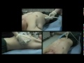
The two available brain imaging systems for this disease are the CT scan, and the MRI brain studies and which is better has been an issue of debate and a lot of studies that we will discuss here.
In most areas of the world, the CT scan brain is still the imaging technique of choice for patients with neurological symptoms due to the fact that it is more readily available and less expensive which gives it a financial advantage over MRI, which is still unavailable in most centers especially in the developing world, also its high cost which is a serious obstacle against its wide use in the acute setting, and makes its use reserved for the chronic conditions in which CT scan studies are not conclusive.
From a medical point of view, the CT scan study is highly informative, and gives a lot of useful data regarding the brain anatomical structures especially with new high resolution multi-slice CT machines. It has a very high sensitivity exceeding 95% in instant detection of brain haemorrhage few minutes after its development, and thus can confirm or exclude the possibility of a haemorrhagic stroke in a reliable way. It can also reveal brain masses and a haemorrhage within a tumour which is sometimes the cause of a sudden deterioration of patients with brain tumours. In expert hands a CT scan study can differentiate between old and acute strokes lesions, and reveal any sort of brain oedema, or atrophic and sclerotic lesions related to aging, and so it is clearly obvious that a CT scan brain study is actually a very useful diagnostic tool, but despite all of these advantages there are 2 major drawbacks that created the whole CT vs. MRI debate.
The first disadvantage is the fact that an ischemic brain stroke may not appear in a CT scan for up to 48 hours period, and thus with a patient presenting with clinical picture suggesting a stroke a CT scan brain can exclude the possibility of a haemorrhagic stroke, but may not confirm the presence of an ischemic lesion for up to 48 hours, which may lead to some therapeutic problems that will be discussed later. The second drawback with CT scanning is the lack of sensitivity to the vertebro-basillar lesions, and taking into consideration that up to 15% of cerebrovascular strokes (either haeomorrhagic or ischemic) are in the vertebro-basillar region, this creates a problem in patients with disturbed level of consciousness and neurological symptoms with an apparently free CT scan study, and necessitates proceeding to MRI to show more anatomical details and reveal any lesion in the vertebro-basillar area.
Having discussed the CT scan imaging drawbacks we can easily understand the points of superiority of the MRI studies. There is no doubt that MRI in simple words is superior to CT scan in the quality of the image and the accuracy of the anatomical details it shows which makes it the imaging modality of choice for patients with gradual chronic neurological symptoms (chronic headache, or gradually developing focal deficits), but in an acute setting the extensive details that MRI gives may not be of importance, but the superiority of MRI depends entirely on the drawbacks of the CT imaging, as an MRI study is very sensitive to ischemic lesions within few minutes of development of an ischemic stroke, and this has a special importance in the new era of thrombolytic therapy for ischemic stroke victims using the tpa agents (tissue plasminogen activator) like alteplase and reteplase, with a very short therapeutic window that is only a three hours period from the beginning of the symptoms and thus requires a solid proof of ischemic stroke immediately which needs a sensitive imaging modality that is the MRI.
It is worth noting that some recent guidelines have recommended applying thrombolytic therapy for patients with a clinical stroke syndrome with brain haemorrhage being excluded by a CT, but despite that recommendation which is based on the lack of availability of MRI in many centers, an MRI study would remain more conclusive and safe, allowing the start of thrombolytic therapy on a solid diagnostic ground.
 The second point of superiority of MRI is its ability to detect the vertebro-basillar lesions which is the second main drawback of the CT scan imaging, but still also some centers for a financial reason applies the "CT scan first" protocol, and proceed to MRI only when a CT scan is not conclusive, which sounds like a reasonable approach from both medical and economic points of view. A third important point of superiority of MRI is in the subarachnoid haemorrhage patient subgroup, especially those with a spontaneous subarachnoid haemorrhage due to an aneurismal rupture, where a surgical clipping or an endovascular coiling of the aneurysm is planned. In such cases an MRI angiography is the imaging modality of choice for assessment of the brain vessels, and determining the aneurismal site and size accurately, allowing an efficient and timely intervention that greatly improves the final outcome in this patient subgroup.
Despite these advantages of MRI there is still an important disadvantage other than the availability and cost issues, which is the few percentage of patients who cannot be imaged with MRI due to claustrophobia, also the patients with cardiac pacemakers, aneurismal metal clips, and any ferro-magnetic materials in their bodies who cannot be safely imaged with MRI. The problem in the acute stroke setting is that many patients who need to be imaged are aphasic or suffering a disturbed level of consciousness and thus cannot give a reliable history on potential contraindications of MRI imaging.
Many studies have been conducted to establish an imaging modality of choice, but the realistic conclusion is that both imaging modalities will co-exist for a long time, as each of them has its advantages and drawbacks in the acute stroke setting, and the imaging modality of choice will always depend on the availability, cost- benefit, and should be tailored to every individual case according to the clinical picture and the other co-morbidities, and after all, a well experienced staff would make the best use of the available diagnostic modality, and effectively guide the therapeutic efforts in the right direction which is the ultimate goal.